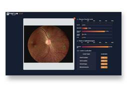
Clinical Image


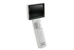
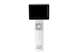
Horus DEC200 Non-Mydriatic Digital Handheld Fundus Camera offers high image quality with ISO 10940 fulfillment.
5MP(2592*1944 pixels)and 45 degree FOV of fundus image are captured to provide more details.
7 internal fixation targets for macula center, disk center and peripheral image in DEC 200 optical modules.
Horus DEC200 provides both auto-focus and power-focus function to facilitate image capturing.
Touch LCD Screen and Wi-Fi compatibility are also equipped .
With a special slit lamp jig, Hours DEC 200 can be mounted with slit lamp for desktop application.


| Lens | |
| Dimension | 201 x 90 x 203 mm |
| Weight | 450 g |
| View Angle | 45 Degree |
| Search Fundus Lighting | natural white/infrared LED |
| Diopter | -20 ~ +20 D |
Q
It's hard to capture a good picture for fundus.
A
Here is the guideline to help you get a good fundus picture by Horus Scope fundus camera.
Q
The image is darker or there are no retinal image available.
A
Reason: The distance or angle for capturing the fundus image is incorrect.
Solution: Please keep the lens aligned with the axis of the eye so that the flash light will enter to retina.
Q
The upper half of the image is clear, but the lower half shows white or blue reflections.
A
Reason: Alignment is ok, but the lens is a little far from the eye.
Solution: Position the lens approximately 2-3 mm close to the eye. The optimal working distance for capturing images is about 24 mm from the front of the lens to the cornea. (The distance may vary slightly depending on different diopter)
Q
1.The fundus image is clear, but the overall picture is a little dark and there is a shadow at the top. 2.The fundus image is clear, but there is a shadow at the top and dazzle on the upper edge.
A
Reason: Exposure Value is not enough or the pupil size is too small.
Solution: 1. Reduce ambient light brightness and fixation light brightness.
2. Increase Exposure Value.
3. Use eye cover of lens, and cover another eye by patient's palm.
4. If the pupil size is below 3.2mm, dilation drops are required.
Q
The upper part of the image is clear, but there is shadowing at the bottom.
A
Reason: The lens was obstructed by the patient's upper eyelid during the capture.
Solution: Let the patient to keep their eyes wide open or hold up their upper eyelid during the capture.
Q
The image is complete but blurry.
A
Reason: The distance and angle betewwn lens and patient's eye are almost correct, but camera or patient doesn't stay stable during photographing.
Solution: Keep camera and patient's eyes steady during photographing.
PS. The Horus fundus camera can be used with a dedicated jig or portable chin rest (CR100).